;)
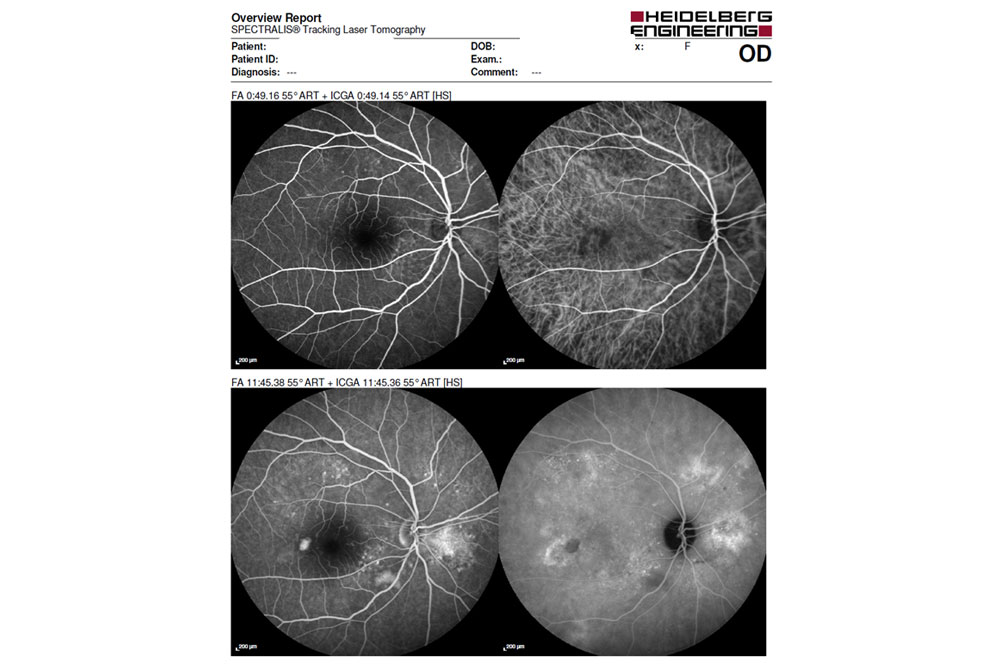;)
Fluorescein absorbs blue light (465-490um) and emits a light of yellow-green spectrum (500-600um). Through the use of specific equipment – the Angiograph, which is nothing more than a photographic apparatus with specific filters, it is possible to serially record the details of the ocular fundus and its vascularization.
By allowing the study of the characteristics of blood flow in the retinal and choroidal vessels, recording details of the pigment epithelium and the retinal circulation and assessing its functional integrity (since normal retinal vessels are impermeable to fluorescein), Fluorescein Angiography is used as an important diagnostic aid in situations of Retinal Vascular Diseases – Diabetic Retinopathy, Arterial Hypertension, Arterial Occlusions and Venous Thrombosis, among others; in Inflammatory or Degenerative situations of the retina and the choroid (age-related Macular Degeneration (DMI) and Retinal Dystrophies), in the study of Ocular Tumors and the Study of the Optic Nerve, and many other primary or non-primary eye diseases.
IMO has the latest generation Angiograph, which requires no “flash”, making it more comfortable for the patient.
IMO has the latest generation Angiograph, which requires no “flash”, making it more comfortable for the patient.
Angiography with Indocyanine Green
It consists of the realization of a set of serialized photographs of the retina under the effect of a contrast agent (Indocyanine Green) administered intravenously.
It evaluates the vascular functioning of the choroid.