Corneal Topography and Tomography of the Anterior Segment
Corneal Topography is based on the projection of a series of concentric rings on the Cornea (Plácido discs), and the reflected image is detected by a video camera. The information collected by measuring the distances between the reflected rings is analyzed by computer software and shown in several ways – tangential, sagittal and three-dimensional colorimetric maps.
More recent imaging tests combine the method described with other technologies, to obtain more information in addition to the study of the anterior surface of the cornea.
The following equipment is available at IMO:


ORBSCAN II
ORBSCAN II combines the information provided by the topography with a slit “scanning” system, which provides elevation maps of the anterior and posterior surface of the cornea, corneal thickness and depth of the anterior chamber.


PENTACAM
PENTACAM is a rotating Scheimpflug camera that performs 50 scans in less than two seconds. It obtains images from the anterior surface of the cornea to the posterior surface of the lens.
This technology constitutes a tomography of the anterior segment, obtaining a real topographic map of the elevation of the anterior and posterior surface of the cornea, corneal aberrometry, map of the real thickness of the cornea (pachymetry), presentation of the image of the whole anterior chamber, allowing for several other measurements, such as depth and volume, to be taken in addition to irido-corneal angle measurement. It also presents the densitometric study of the lens, providing information on the evolutionary state of the cataracts, through the PNS (Pentacam Nucleus Stage) index.
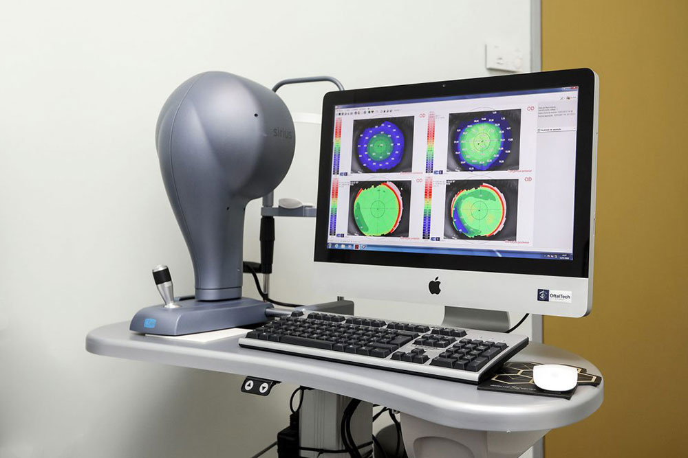
SIRIUS CSO
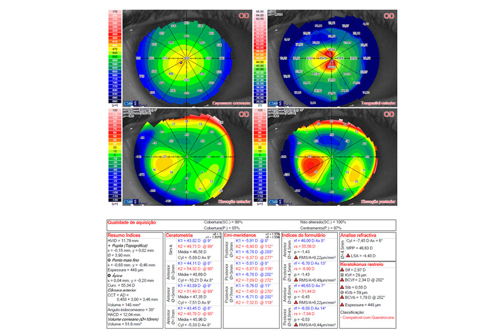
SIRIUS CSO is also a Scheimpflug camera providing additional information, such as the pupillometric study, in different lighting environments. With this equipment we obtain topography, aberrometry and anterior elevation map of the cornea, for the personalized treatments with the Laser Excimer Schwind Amaris.
All these devices have formulas for the detection of subclinical keratoconus, which is essential for all candidates for corneal refractive surgery with the Laser Excimer.